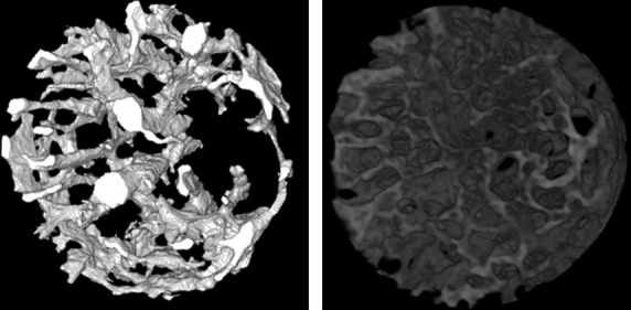
Figure 2.

Three-dimensional displays of blood vessels (left) and bone (right) in a cylindrical region for analysis (diameter 720 μm, height 300 μm) from the cortical defect in Fig.1, produced by SRSμCT. Bones were segmented as extravascular regions with the voxel intensity corresponding to d.HAp > 500 mg/cm3.
