Figure 5.

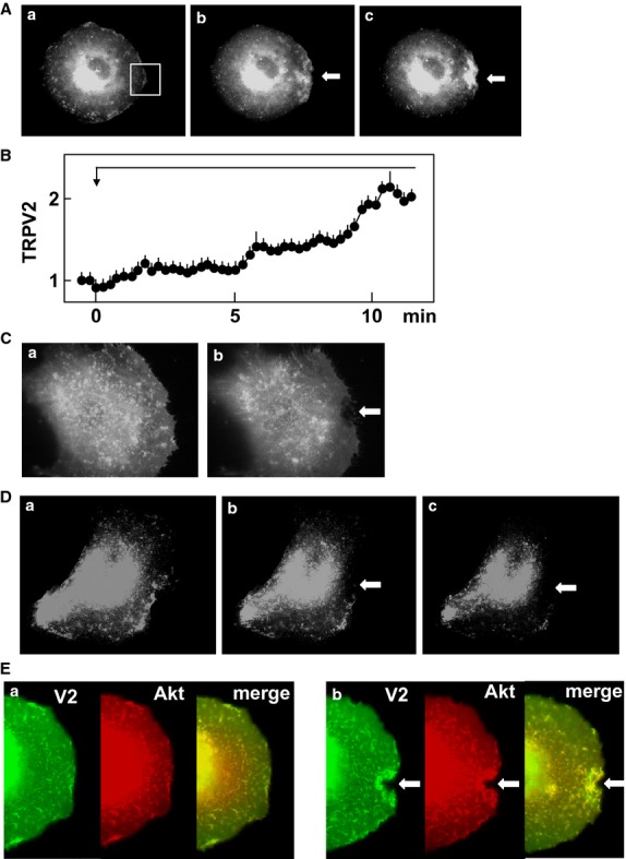
Effect of focal application of mechanical stress on localization of TRPV2. (A) Effect of focal mechanical stress on localization of TRPV2. Mechanical stress was applied at the site indicated by the arrow. Localization of GFP-TRPV2 is shown. a: before the application, b: 1 min, c: 5 min after the application. The results are the representative of more than 50 experiments. (B) Time course of accumulation of TRPV2 beneath the pipette. Mechanical stress was applied to the cells as shown in A. Changes in the intensity of fluorescence of GFP-TRPV2 in area shown in Fig.5A-a were monitored. Values are the mean ± SE for five experiments. (C) Effect of mechanical stress in the absence of extracellular calcium. Mechanical stress was applied as shown in A in the absence of extracellular calcium. The results are the representative of two experiments. a: before application of mechanical stress, b: 5 min after the application of mechanical stress. (D) Effect of LY29034 on translocation of TRPV2. Focal mechanical stress was applied as indicated above in the presence of 100 μmol/L LY29034. The results are the representative of five experiments. a: before the application, b: 1 min, c: 5 min after the application. (E) Effect focal application of mechanical stress on localization of TRPV2 and Akt. Focal mechanical stress was applied as indicated above and localization of GFP-TRPV2 (green) and RFP-Akt (red) was monitored. The results are the representative of four experiments. a: before the application, b: 5 min after the application.
