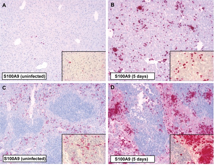
Fig 3. Immunostaining for S100A9 in the liver and spleen of mice during S. Typhimurium infection.
Positive immunostaining for S100A9 in liver parenchyma (A) and the red pulp of the spleen (C) tissue was observed in uninfected control animals. Five days after infection with S. Typhimurium (106) there was a marked increase of S100A9 in both liver (B) and spleen (D) corresponding with inflammatory cell influx. Magnification 10 × and 40 ×.

