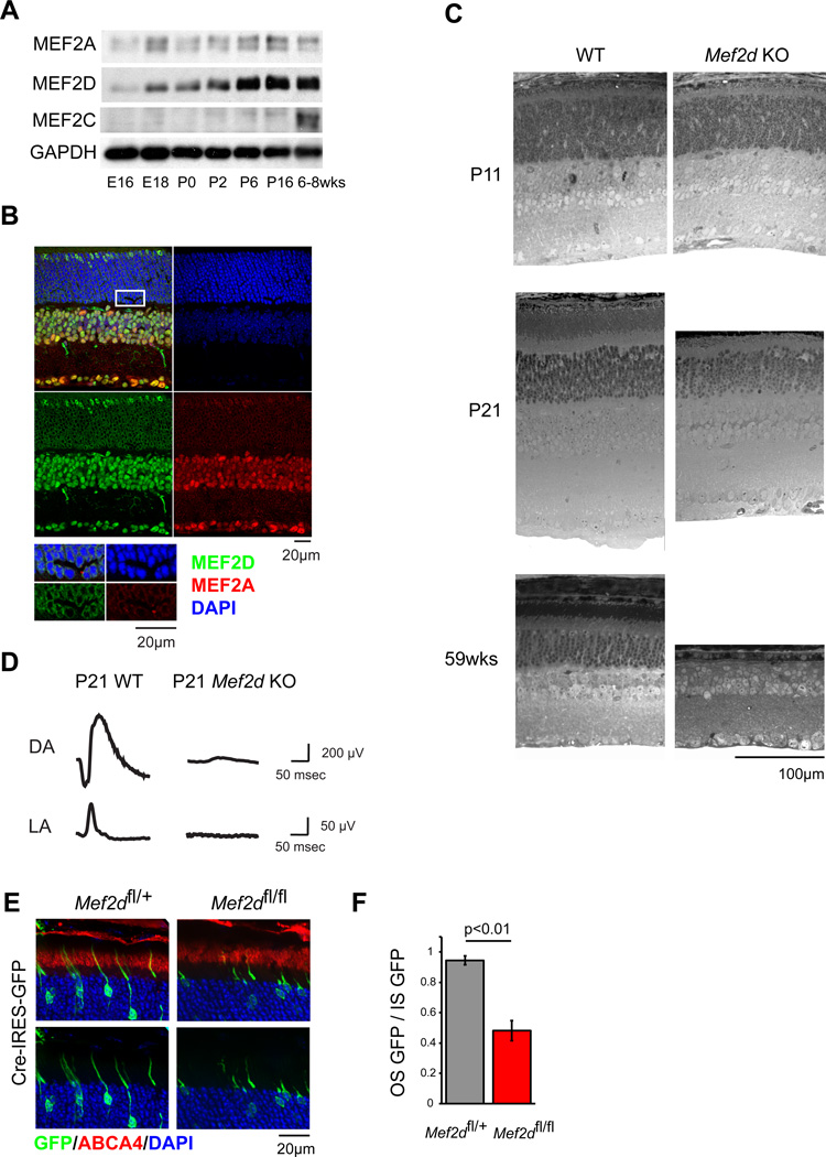
Figure 1. MEF2D is required cell-autonomously for photoreceptor development and function.
(A) Immunoblot of MEF2 family member expression in mouse retina over development. (B) Immunofluorescence of P25 WT retinal cross-sections for MEF2A (red) and MEF2D (green) with DAPI (blue). Inset highlights rod photoreceptor nuclei. ONL, outer nuclear layer; INL, inner nuclear layer; GCL ganglion cell layer. (C) Toluidine blue-stained cross-sections of WT and Mef2d KO littermate retinas. OS, photoreceptor outer segments; IS, photoreceptor inner segments. (D) Representative electroretinograms (ERGs) from a P21 WT mouse and a Mef2d KO littermate under dark-adapted (DA) and light-adapted (LA) conditions. (E) Representative immunofluorescence images of P21 retinas electroporated at P0 for sparse expression of Cre recombinase and GFP (green) in either Mef2dfl/+ or Mef2dfl/fl mice. ABCA4 immunostaining (red) was used to identify photoreceptor cell outer segments (OS). (F) Quantification of OS from GFP-positive photoreceptors as shown in (E). Mean GFP intensity in the ABCA4-positive region (OS GFP) was normalized to mean GFP intensity in the inner segments (IS GFP) to measure OS development while controlling for electroporation density (Mef2dfl/+, N=6; Mef2dfl/fl, N=3 retinas). Error bars represent S.E.M.

