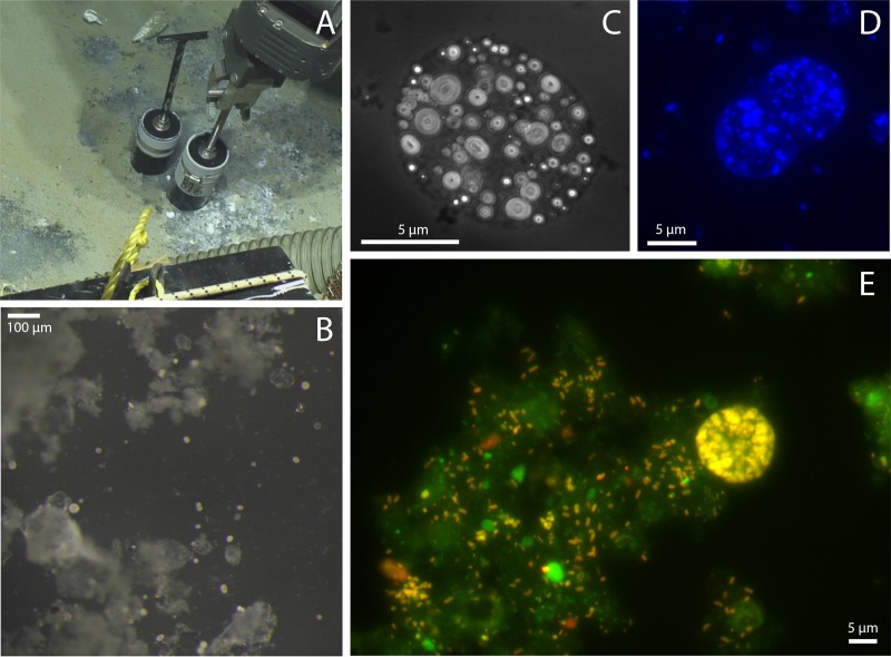
FIG 2.
Images of cold seeps and Barbados “Ca. Thiopilula” cells. (A) Push core collection of cold seep sediments at a 4,743-m water depth. (B) Low-magnification image of the white biomat in panel A showing large spherical cells (5 to 40 μm in diameter) in the biofilm. (C to E) Photomicrographs of “Ca. Thiopilula” cells and associated microorganisms in the biofilms. (C) Phase-contrast image showing intracellular sulfur inclusions; (D) DAPI-stained cells; (E) FISH photomicrograph of postincubation sediments using the general bacterial probe EUBMIX with both 6-carboxyfluorescein (green) and cy5.5 (red) filter sets.

