Fig. 2.

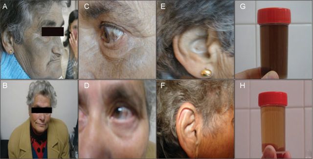
Ochronotic pigmentation of skin, sclera, ear cartilage and urine in Patient 1 (A, C, E and G, respectively) and Patient 2 (B, D, F and H, respectively). Note enhanced pigmentation in Patient 1.

Ochronotic pigmentation of skin, sclera, ear cartilage and urine in Patient 1 (A, C, E and G, respectively) and Patient 2 (B, D, F and H, respectively). Note enhanced pigmentation in Patient 1.