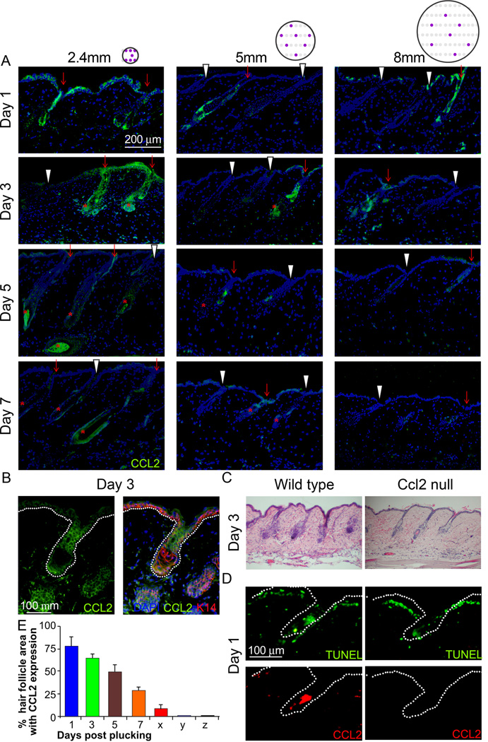
Figure 4. CCL2 is involved in plucking induced hair regeneration.
(A) HF keratinocytes showed higher CCL2 expression (green) in plucked follicles (red arrow) than in unplucked follicles (white arrowhead). The circle with purple dots indicates the topology of plucked follicles (please also see Fig. 1E). Peak expression occurs 1–3 days after plucking, and no marked difference between the 2.4, 5 and 8mm groups were noted. (*: Regenerating HFs) (B) Double immunostaining for K14 and CCL2 of samples 3 days after plucking showed that HF keratinocytes in plucked follicles are the main source of CCL2. (C) Hair re-growth is retarded when hairs were plucked from CCL2 null mice. (D) CCL2 null mice showed similar apoptotic HF cells following plucking as wild type mice, but couldn’t induce CCL2 in apoptotic HF cells. (E) Graph showing the percentage of HF area expressing CCL2 at 1, 3, 5 and 7 days post plucking as well as unplucked HFs within (x) and outside (y) of the plucked field. CCL2 null mice do not express CCL2 (z). (n=3). (Data are represented as mean +/− S.D.). See also Figure S5.

