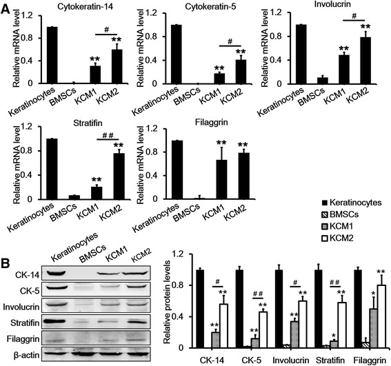
Figure 6.

Quantitative real-time-polymerase chain reaction and Western blot analysis of keratinocyte markers after a 21-day treatment with two different keratinocyte-conditioned media. (A) mRNA expression of cytokeratin-5, cytokeratin-14, stratifin, involucrin, and filaggrin in bone marrow mesenchymal stem cells (BMSCs), keratinocytes, and KCM1 and KCM2 groups. Keratinocytes at passages 2 to 5 and BMSCs at passages 4 to 7 were used as positive and negative controls, respectively. Keratinocytes were normalised to 1 because these cells expressed all of the markers. (B) Western blot analysis of cytokeratin-5, cytokeratin-14, stratifin, involucrin, and filaggrin in BMSCs, keratinocytes, and KCM1 and KCM2 groups. Keratinocytes at passages 2 to 5 and BMSCs at passages 4 to 7 were used as positive and negative controls, respectively. Beta-actin was used as a loading control. Data are presented as the mean ± standard error of the mean. n = 3. *P <0.05, **P <0.01 BMSCs versus KCM1/KCM2, # P <0.05, ## P <0.01 KCM1 versus KCM2. CK-5, cytokeratin-5; CK-14, cytokeratin-14; KCM1, keratinocyte-conditioned medium collected from keratinocytes with no Y-27632 treatment; KCM2, keratinocyte-conditioned medium collected from keratinocytes treated with Y-27632.
