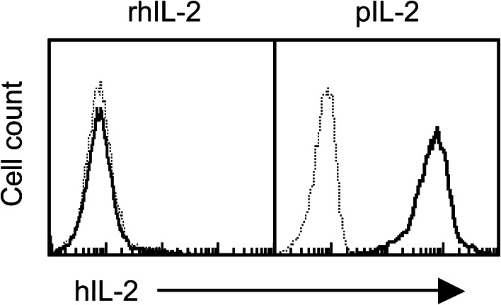
Fig. 1.
Protein transfer (painting) of pIL-2 onto T cells. OT-1 CD8+ T cells were incubated with pIL-2 or rhIL-2 (30 μg/ml/107 cells) at 37 °C for 10 min, washed, stained with biotinylated rabbit antihIL-2 Ab and APC streptavidin, and analyzed by flow cytometry. Dotted line, non-transferred cells; solid line, transferred cells. Shown is 1 of 3 experiments with consistent results. Incubation with palmitate-derivatized control proteins did not result in positive staining for IL-2 (not shown)

