
Fig 2.
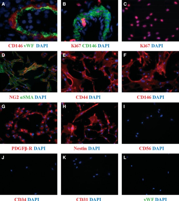
Immunofluorescence of human pericytes before and after long-term culture. Immunohistochemistry on frozen section of adult pancreas (a) and foetal skeletal muscle (b) and immunocytochemistry on cultured pericytes (c–l) show the expression of CD146, NG2, αSMA, CD44, PDGFRβ and nestin by pericytes before and after culture. Endothelial (vWF, CD34, CD31) and neural and myogenic (CD56) markers are absent. Few pericytes express Ki67, marker of proliferation, in situ (b) compare to cultured pericytes (c). All nuclei are stained by DAPI (blue). Pericyte culture and immunostainings were performed according to our established protocol and human developing and adult tissues were used according to University of Pittsburgh regulations 21. Magnifications: 600× (a); 400× (b) and 200× (c–l).