
Fig 4.
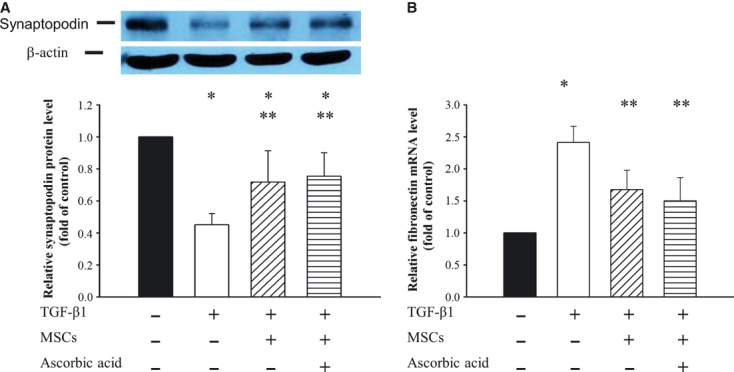
Effects of conditioned MSCs on expression of synaptopodin and fibronectin in podocytes. Podocytes were incubated without or with 15 ng/ml TGF-β1, conditioned MSCs and/or ascorbic acid for 72 hrs. Cell lysates were analysed with Western blotting. (A) Representative Western blot and bar graph analysis of relative protein level of synaptopodin, which were normalized to control. (B) Bar graph analysis of quantitative PCR analysis of relative fibronectin expression to GAPDH normalized to control. *P < 0.05 as compared with control; **P < 0.05 versus TGF-β1 treated.