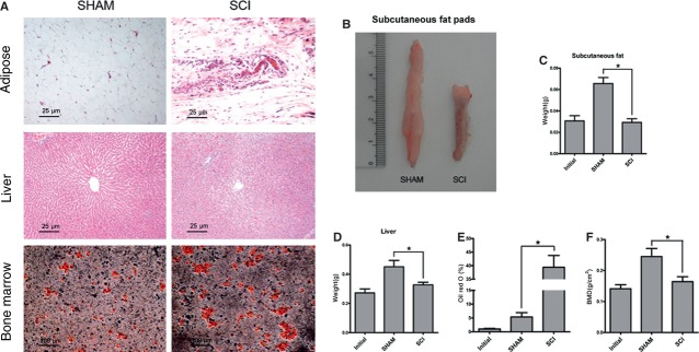
Fig 1.

Adiposity is increased in tibial bone marrow, in contrast with peripheral adipose tissue lipolysis. (A) Representative fat pad sections, liver sections stained with haematoxylin and eosin, and bone marrow sections stained with Oil red O from spinal cord injury (SCI) and SHAM rats. Adipose tissue obtained from femoral fat pads and the liver of SCI rats exhibited lipid sparse adipocytes compared with that of SHAM rats. Bone marrow from SCI rats stained for oil red O displayed a significant increase in adipocytes compared with that of SHAM rats. (B) Representative subcutaneous fat pads from SCI and SHAM rats. (C) The values of weights of subcutaneous fat pads were pooled from 10 rats per group and expressed as averages ± SE. *P < 0.01. (D) The values of weights of livers were pooled from 10 rats per group and expressed as averages ± SE. *P < 0.01. (E) The values of percentage of Oil red O staining were pooled from 10 rats per group and expressed as averages ± SE. *P < 0.01.
