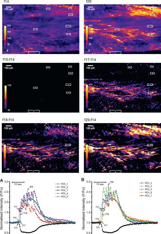
Fig 4.

Propagation of [Ca2+]i transient in myometrial slice containing two bundles of myocytes, one in the middle, another in the top right corner. Data presentation is similar to that in Figure3 with the exception that two sets of three ROIs were used in this experiment: ROI 1-2 and ROI 4-5 to measure propagation perpendicular to the longitudinal axis in each bundle and ROI 2-3 and ROI5-6 to measure propagation in axial direction. In this experiment, there was a 1 sec. time delay between the onset of [Ca2+]i transients in neighbouring bundles but cells within each bundle were activated simultaneously. Typical of seven experiments on slices from seven different samples.
