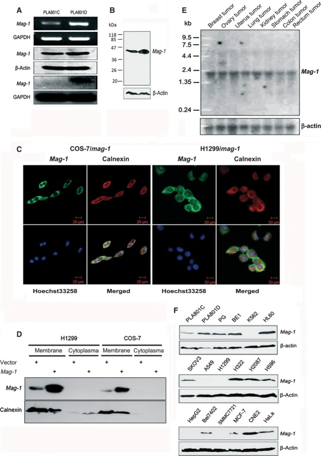
Fig 1.

Identification and expression profile of mag-1 gene. (A) Verification of differential expression of mag-1 in PLA801C and D cells. Upper two lines indicate semi-quantitative RT-PCR analysis of mag-1 mRNAs. Middle two lines are Western blot analysis of Mag-1 protein levels using β-actin as internal control between PLA801C and D. Lower two lines indicate northern blot analysis of mag-1 mRNAs using GAPDH as internal control between PLA801C and PLA801D. (B) Molecular weight of Mag-1 protein analysed using Western blot analysis. (C) Subcellular localization of Mag-1 within cells. A immunofluorescent staining of COS-7 and H1299 cells overexpressing His-Tagged Mag-1. Mag-1 was stained with anti-His antibody (Green) and visualized co-localization with ER specific protein, Calnexin (Red). Cells were double stained with Hoechst 33258 (blue) to identify the nuclei in the corresponding fields. All cell samples were visualized using Confocal laser-scanning microscope (Zeiss 510 META, Oberkochen, Germany). (D) Western blot analysis of cell membrane and cytoplasma fraction from H1299 and COS-7 cells, stably transfected mag-1 or vector control. (E) Northern blot analysis of mag-1 transcription in eight human tissues. A human multiple tissue northern blot membrane was hybridized with 32P-labelled mag-1 specific or β-actin cDNA probes (bottom) respectively. Hybridization with β-actin served as a loading control. Size marker is indicated on the left. (F) Western blot analysis of mag-1 protein in multiple cancer cell lines. β-Actin was used as an internal control.
