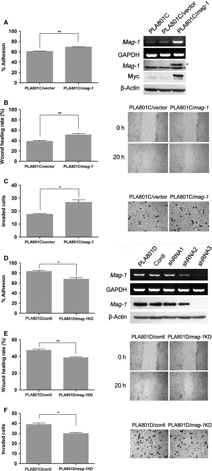
Fig 2.

Effect of mag-1 on cell adhesion, migration and invasion capability of human lung cancer cells in vitro. (A) Adhesion of PLA801C/mag-1 and mock control cells to Matrigel. The cells were added to each 96-well coated with Matrigel. After 1 hr of incubation, tumour cell adhesion was measured using MTT assay. Results were shown as the adhesion rate calculated according to the formula: (A490 nm of the adhered cells/A490 nm of total cells)×100%. The exogenous mag-1 expression was analysed by RT-PCR (upper two lines) and Western blot assay (middle two lines). Arrow indicates myc-tagged Mag-1 protein. (B) Cell migration after injury. A total of 7 × 10⁵ cells per well were seeded on a 6-well plate in normal culture medium and allowed to reach 100% confluence. The injury line was introduced to scratch the cell monolayer with a sterile pipette tip of 2 mm in width, and then cells were rinsed with PBS and incubated for 20 hrs. Wound closure was monitored by visual examination using an Olympus microscope. (C) Transwell cell invasion assay. 1 × 10⁵ cells were plated in the upper layer of each transwell (coated with 120 μg basement membrane, Matrigel) and then, incubated at 37°C for 24 hrs. After incubation, the non-migrating cells were removed from the upper surface of the membrane and the penetrated cells on the lower surface of the membrane were stained with H.E. Results were plotted as the average number of cells of three random visual fields that migrated through the membrane. (D) (right) Effect of mag-1-targeted shRNA constructs and scramble control on mag-1 expression in PLA801D cells. Transfected cells were selected with G418 for 3 weeks until stable clones were obtained and expanded for other experiments. The mRNA and protein level of mag-1 were assayed using RT-PCR and Western blot. A(left) Adhesion of PLA801D/shRNA and control cells to Matrigel. (E) Wound healing migration assay and (F) transwell cell invasion assay. The shRNA and scramble control stable transfected cells were cultured and measured using the same methods as mentioned before. Each assay was performed thrice in triplicate. In all studies, statistical significance was determined using One-way anova with non-parametric analysis comparing data points to control. Bars show the standard error of the mean. *P < 0.05; **P < 0.01.
