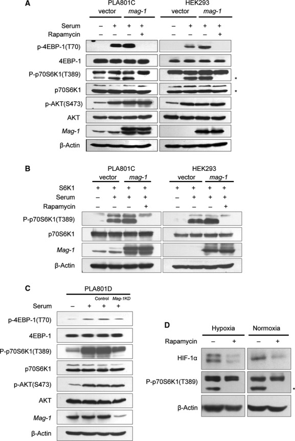
Fig 6.

mag-1 induces activation of mTOR signalling transduction pathway. (A) HEK293 and PLA801C cells were transfected with pcDNA3.1/mag-1 and control vector for about 12 hrs followed by serum starvation for 24 hrs and then stimulated with 10% foetal bovine serum for 20 min. prior to cell lysis. For inhibitory assay, cells were treated with 20 nM rapamycin 30 min. before serum stimulation. The levels of regulators in PI3K/AKT/mTOR signalling pathways were detected using Western blot using the antibodies shown to the left of the panels. (B) PLA801C and HEK293 cells were co-transfected with pcDNA3.1/mag-1 and pcDNA3.1/S6K1 plasmids followed by the treatment as mentioned above. The phosphorylation of exogenous p70S6K1 (T389) was analysed using Western blot. (C) PLA801D cells stably transfected with mag-1/shRNA or scramble control shRNA were cultured with serum-free medium for 24 hrs and then stimulated with 10% foetal bovine serum for 20 min. prior to cell lysis. The phosphorylation of p70S6K1 (T389), 4EBP-1(T70) and AKT (S473) were analysed using Western blot using the specific antibodies respectively. (D) H1299/mag-1 cells were exposure to hypoxia for 8 hrs or under normoxia, followed by 20 nM rapamycin treatment for 1 hr prior to cell lysis. The level of HIF-1α and phosphorylation of p70S6K1 (T389) was analysed using Western blot. The line of β-Actin indicated the equal loading as an internal control.
