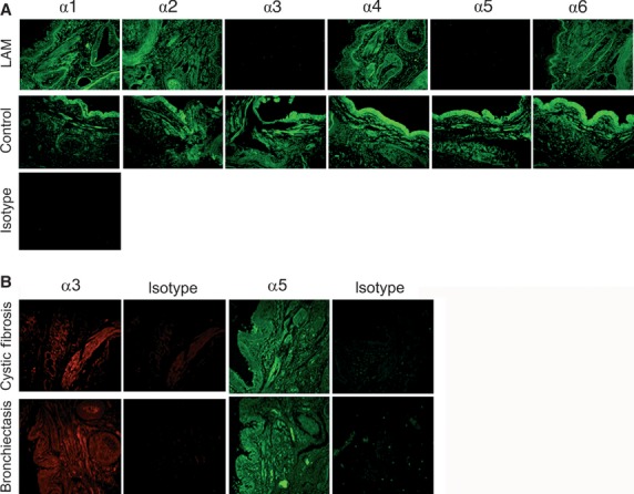
Fig 1.

Immunohistochemical staining for Col IV NC1 isoforms. (A) Col IV NC1 domains α1–α6 were stained in human lung tissue sections using specific antibodies and detected using a secondary antibody conjugated to fluorescein isothiocyanate (FITC) (green staining). Eight lymphangioleiomyomatosis (LAM) patients and 10 controls were tested. Scale bar represents 50 μm. L: lumen; B: basal lamina; V: blood vessel. (B) Col IV α3 (red staining) and α5 (green staining) NC1 domains were detected in human lung tissue sections from four cystic fibrosis patients and one bronchiectasis patient. The isotype control was exposed to rat IgG and the secondary antibody.
