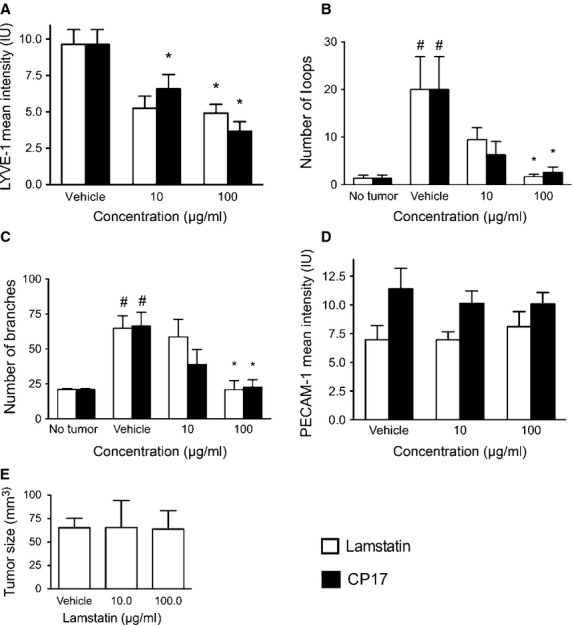
Fig 6.

Lamstatin and CP17 inhibit lymphangiogenesis in a murine model of tumour-induced lymphangiogenesis. LNM35 tumour cells, expressing EGFP, were injected intradermally into the ears of NOD/SCID/gamma mice with or without lamstatin (white bars) or CP17 (black bars) and Matrigel. (A) Mean intensity values of LYVE-1 fluorescence staining in lamstatin and CP17 treated animals (Lamstatin vehicle n = 11, 10 and 100 μg/ml lamstatin n = 8 and n = 9, CP17 vehicle n = 11, 10 and 100 μg/ml CP17 n = 8 and n = 6, mice respectively). *P < 0.05 one-way anova (Kruskal–Wallis, with Dunn's multiple comparisons corrections) treatment versus vehicle. *P < 0.05. (B) Assessment of loops in tumour ear model of lamstatin-treated animals (no tumour n = 3, vehicle n = 11, 10 and 100 μg/ml lamstatin, n = 8 and n = 9 mice respectively) and CP17-treated animals (no tumour n = 3, vehicle n = 11, 10 and 100 μg/ml CP17, n = 8 and n = 6 mice respectively). (C) Morphological analysis of branching points of lymphatic vessels in whole mount staining of LNM35 tumours with lamstatin (no tumour n = 3, vehicle n = 11, 10 and 100 μg/ml lamstatin, n = 8 and n = 9 mice respectively) or CP17 (no tumour n = 3, vehicle n = 11, 10 and 100 μg/ml CP17, n = 8 and n = 6 mice respectively) treatment; (D) Percentage of mean PECAM-1-positive blood vessel area (% mean PECAM-1 PVA) of animals treated with vehicle, lamstatin (vehicle (292.2 ng/l EDTA, pH 3.5) n = 11, 10 and 100 μg/ml lamstatin n = 8 and n = 9 mice respectively) or CP17 (vehicle (MilliQ water) n = 11, 10 and 100 μg/ml CP17 n = 8 and n = 6 mice respectively). All data presented as mean ± SEM, one-way anova (Kruskal–Wallis, with Dunn's multiple comparisons corrections) treatment versus no treatment, *P < 0.05. (E) Lamstatin has no effect on tumour size after 12 days. After 12 days tumour sizes were measured in animals treated with either vehicle (292.2 ng/l EDTA, pH 3.5), 10 or 100 μg/ml lamstatin. Data are expressed as mean ± SEM. Vehicle n = 11, 10 and 100 μg/ml lamstatin n = 8 and n = 9 mice.
