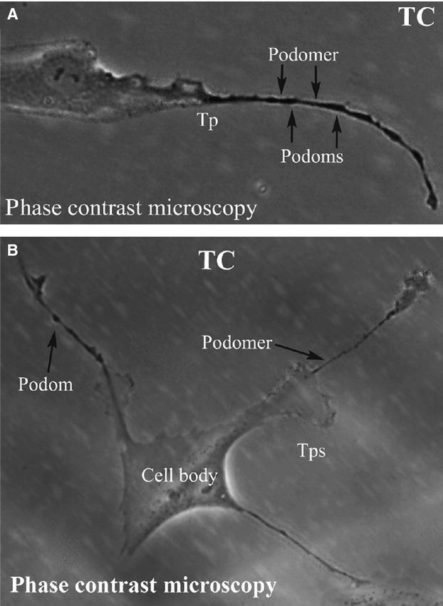
Fig 1.

(A) and (B) Phase contrast microscopy of kidney TCs in primary culture. Note the typically very long Tps (over 50 μm). Also, the specific structure of Tps is obvious: alternation of dilations (podoms) with thin segments (podomers). Direct magnification: 400×. TC: Telocyte; Tp: Telopode.
