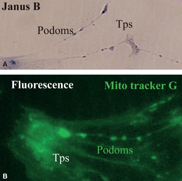
Fig 2.

(A) and (B) Kidney TCs in primary culture. (A) Vital staining for mitochondria with Janus Green B; note the podoms coloured blue-brown because of the presence of mitochondria. (B) Fluorescence microscopy using Mito Tracker Green as molecular probe for the presence of mitochondria; note the moniliform aspect of Tps as a result of accumulation of mitochondria in podoms. TC: Telocyte; Tp: Telopode; Mito Tracker G: Mito Tracker Green.
