
Figure 1. Schematic diagram illustrating hyaluronan biosynthesis.
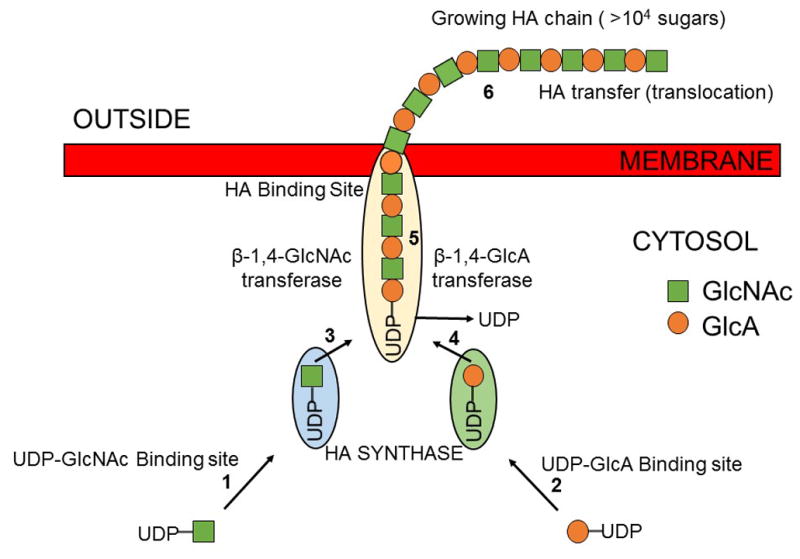
A membrane-bound hyaluronan synthase utilizes uridine diphospho-glucuronic acid (UDP-GlcA) and uridine diphospho-N-acetylglucosamine (UDP-GlcNAc) as substrates for hyaluronan biosynthesis. The UDP-sugar substrates are used by the hyaluronan synthase inside the cell and the hyaluronan chain is continuously translocated across the membrane at the reducing end. Two enzymes are shown to illustrate how one enzyme could alternately make each of the two types of glycoside bonds in hyaluronan.