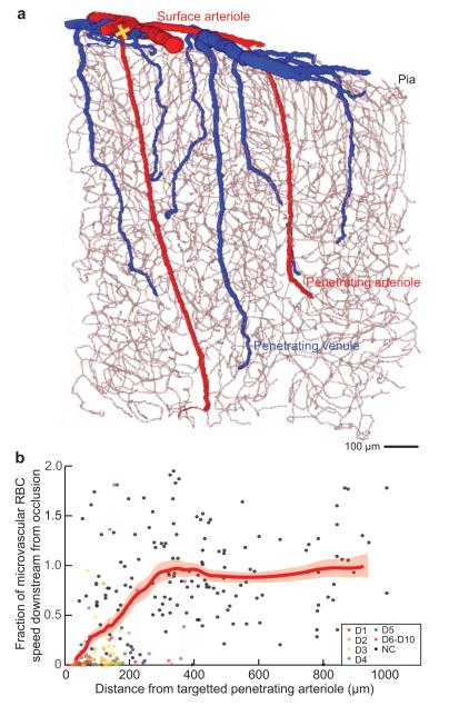
Figure 5. The penetrating arterioles are a fragile link in the delivery of blood from the surface arterioles to the subsurface microvascular network.
(a) Numerical section of a vectorized network from a complete reconstruction (Figure. 1) that highlights the penetrating arterioles (red) and venules (blue) and the continuous microvascular network (gray). Adapted from Blinder et al. [17]. (b) The fraction of speed in neighboring microvessels that lie in cortical layers 2/3 after an occlusion to a single microvessel, relative to that before the occlusion. Only some of the data points could be specified in terms of downstream branch number, as shown. The thick red line through the data is the smoothed response averaged over a window that included 50 points, with ± 1 SEM limits indicated by the thin red lines. The data is averaged over 16 networks. Adapted from Nishimura et al. [23].

