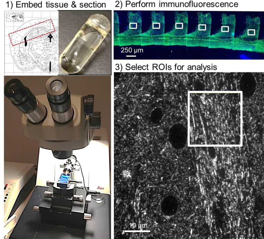
Figure 1. Array tomography workflow.
(1) Tissue is cut into 5 × 1.5 × 1 mm blocks containing corpus callosum and external capsule (Franklin and Paxinos, 2004) which are embedded in LR white media in gelatin capsules (inset, top). An ultramicrotome is used to produce 70–90 nm section ribbons using a histojumbo diamond knife. (2) Standard immunofluorescent techniques are used to label each ribbon and images from sections are captured using a 63× lens on an epifluorescent microscope. (3) Each image can then be further subdivided into smaller regions for analysis, excluding cell bodies and tissue processing artifacts.

