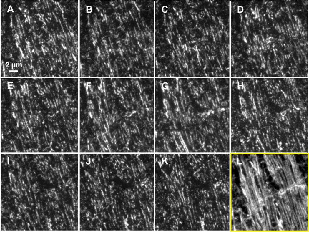
Figure 2. Example of a short array containing uninjured mouse external capsule labeled with anti-tubulin and Alexa 488.
(A–K) Images of eleven 70 nm thick ultrathin sections labeled with anti-tubulin and Alexa 488. Images have been co-registered so that each represents the same 19.5 × 19.5 µm area. (L) A projection of the 11 image stack shows a reconstruction of individual, longitudinally/transversely cut axons within this stack.

