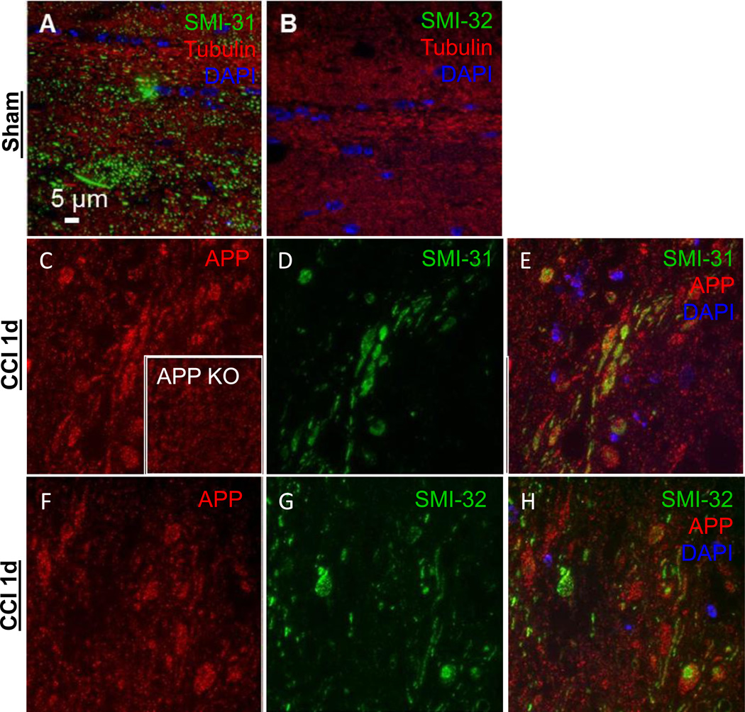
Figure 5. Axonal injury markers SMI-31, SMI-32 and APP 24 hours in uninjured sham or 1.0 mm CCI in the ipsilateral external capsule.
(A) SMI-31-Alexa 488 labels axons in an uninjured mouse while (B) SMI-32-Alexa 488 does not. Both images are co-labeled for tubulin- Alexa 594 to visualize axons and DAPI to indicate cell nuclei. After injury, (C) APP Cy3 labeled axons (inset is from CCI injured APP knockout mouse indicating non-specific binding). (D) SMI- 31 and (E) composite image of DAPI, SMI-31, and APP. (F) APP Cy3 labeled axons. (G) SMI-32 Alexa 488 labeled axons. (H) Composite image of DAPI, SMI-32, and APP. All images are from wild-type mice except panel C inset.

