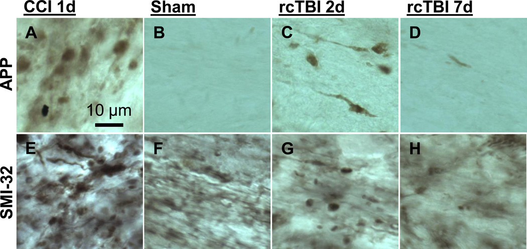
Figure 6. Conventional immunohistochemistry for APP and SMI-32 in external capsule.
Representative images of axonal injury from 1 day CCI (A, E), 7 day sham (B, F), 2 day rcTBI (C, G) or 7 day rcTBI (D, H) in wild-type mice. Sections were labeled for amyloid precursor protein (A, B, C, D) or with antibodies to SMI-32 (E, F, G, H). Images in CCI mice were captured from pericontusional corpus callosum. All other images were taken from external capsule directly underlying the site of impact (or sham injury) near the lateral ventricle, which was the only region where axonal varicosities were observed using these markers.

