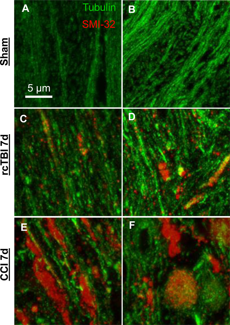
Figure 7. Projection images of arrays (~20 sections each) from external capsule labeled with the axonal injury marker SMI-32-Alexa 594 and tubulin-Alexa 488.
(A, B) Axons from uninjured wild-type mice displayed little SMI-32 labeling. (C,D) Mice subjected to repetitive concussive traumatic brain injury had punctate areas of SMI-32 labeling and occasional colocalization of SMI-32 and tubulin in swollen axons at 7 days. (E,F) Larger axonal varicosities >3 µm in diameter were apparent in a model of 1.5 mm CCI moderate traumatic brain injury at 7 days. Tubulin loss was also evident. Panels in each row represent images of arrays from two separate mice.

