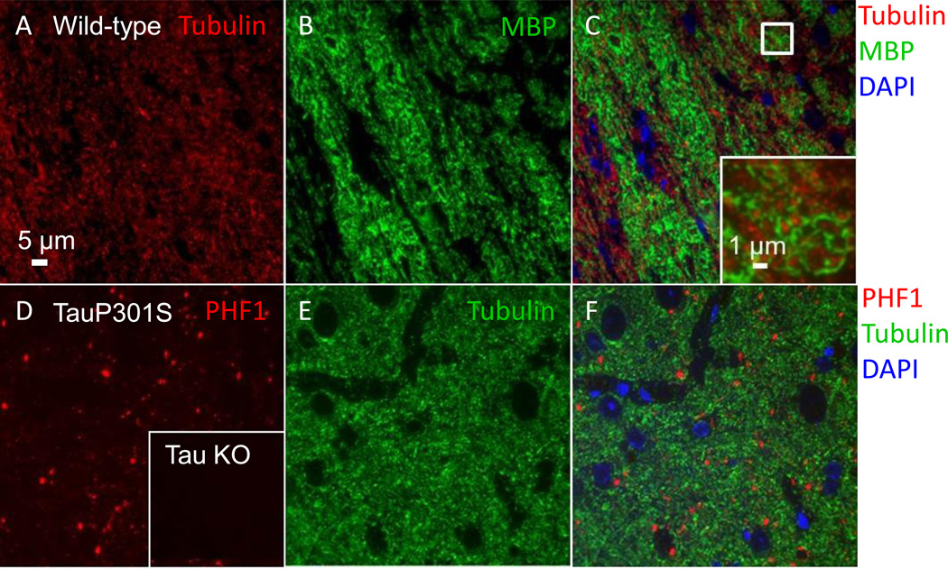
Figure 9. Additional axonal markers for use with array tomography.
(A–C) Myelin basic protein and tubulin labeling in the external capsule of an uninjured wild-type mouse. (A) Tubulin- Alexa 594 labeled axons. (B) Myelin basic protein (MBP)-Alexa 488 labeled axons. (C) Composite image of DAPI, myelin basic protein, and tubulin. Inset shows an enlarged view of the box in (C), where individual myelinated axonal cross-sections are clearly visible. (D–F) PHF-1 tau and tubulin labeling in the entorhinal cortex of a 12-month-old Tau P301S mouse. (D) PHF1-Cy3 labeled punctae. Inset shows the absence of PHF1 labeling in cortex from a tau knockout mouse. (E) Tubulin Alexa 488 labeled neuropil. (F) Composite image of DAPI, tubulin, and PHF1.

