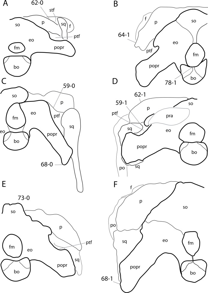
Figure 14. Sauropod skulls in posterior view.
Sauropod skulls of Spinophorosaurus nigerensis GCP-CV-4229 (A; traced from Knoll et al., 2012); Suuwassea emilieae ANS 21122 (B; traced from Harris, 2006a); Limaysaurus tessonei MUCPv-205 (C; after Calvo & Salgado, 1995); Kaatedocus siberi SMA 0004 (D); Apatosaurus louisae CM 11162, (E, reversed); Diplodocus sp. CM 11161 (F) in posterior view. Note the participation (C; C59-0) or exclusion (D; C59-1) of the parietal to the posttemporal fenestra; the straight (A; C62-0) or convex (D; C62-1) dorsal edge of the posterolateral process of the parietal; the outwards curve of the distal end of the posterolateral process of the parietal (B; C64-1); the distally expanded (C; C68-0) or straight paroccipital processes (F; C68-1); the dorsally vaulted supraoccipital (E; C73-0); and the narrow contribution of the basioccipital to the dorsal surface of the condyle (B; C78-1). Skulls scaled to the same occipital condyle width.

