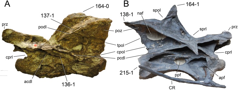
Figure 41. Cervical vertebra 6 of neosauropods.
Cervical vertebra 6 of Australodocus bohetii MB.R.2455 (A) and Galeamopus sp. SMA 0011 (B) in left (A) and right (B) lateral view. Note the short second pcdl in Australodocus (A; C136-1), the foramen piercing the podl (A; C137-1), the projection formed by the epipophysis (B; C138-1), the low (A; C164-0), and high (B; C164-1) neural spines, and the cervical rib, which is slightly longer than the centrum in Galeamopus (B; C215-1). Abb.: acdl, anterior centrodiapophyseal lamina; apf, anterior pneumatic fossa; cpol, centropostzygapophyseal lamina; cprl, centroprezygapophyseal lamina; CR, cervical rib; naf, neural arch foramen; pcdl, posterior centrodiapophyseal lamina; podl, postzygodiapophyseal lamina; poz, postzygapophysis; ppf, posterior pneumatic fossa; prz, prezygapophysis; spol, spinopostzygapophyseal lamina; sprl, spinoprezygapophyseal lamina; tpol, interpostzygapophyseal lamina. Vertebrae scaled to the same centrum length.

