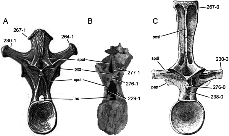
Figure 63. Diplodocoid posterior dorsal vertebrae.
Posterior dorsal vertebrae of Haplocanthosaurus priscus CM 572 (A; modified from Hatcher, 1903), Demandasaurus darwini MDS-RVII 798 (B; modified from Torcida Fernández-Baldor et al., 2011), and Apatosaurus louisae CM 3018 (C; modified from Gilmore, 1936) in posterior view. Note the paired pneumatic foramen dorsolateral to the neural canal in Demandasaurus (B; C229-1), the different orientations of the diapophyses in Haplocanthosaurus (A; C230-1) and Apatosaurus (C; C230-0), the single lamina that supports the hyposphene from below (C; C238-0), the dorsal spur on the tip of the transverse process (A; C264-1), the small triangular lateral projections at the spine top (A; C267-1), or their absence (C; C267-0), the rhomboid (C; C276-0) in contrast to laminar (B; C276-1) hyposphene, and the ventrally forked spol (B; C277-1). Abb.: cpol, centropostzygapophyseal lamina; nc, neural canal; pap, parapophysis; posl, postspinal lamina; poz, postzygapophysis; spdl, spinodiapophyseal lamina; spol, spinopostzygapophyseal lamina. Scaled to same posterior centrum height.

