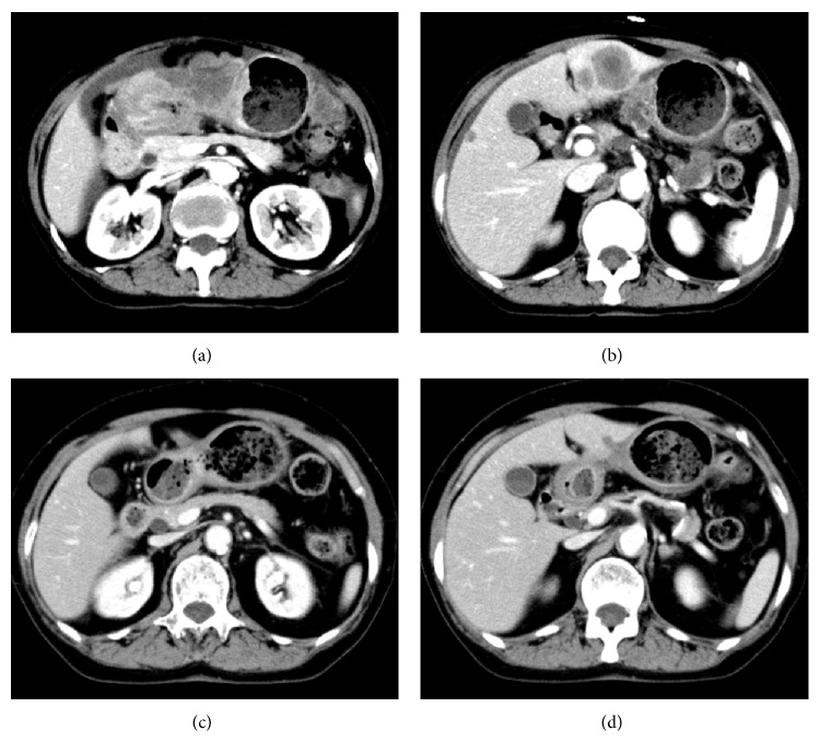
Figure 3.
Contrast-enhanced computed tomography (CT) findings. (a) and (b) show the CT at presentation. (a) The wall of the lower part of the stomach is markedly thickened, and the density of the surrounding fat tissue is increased, forming an 80 × 45 mm mass. (b) Two nodules are detected in the lateral segment of the liver, and these are directly contiguous from the gastric mass. (c) and (d) show the CT findings after completion of the 11th course of chemotherapy (capecitabine + CDDP + trastuzumab). (c) The gastric mass is markedly decreased. (d) Only a small nodule in the lateral segment of the liver is detected.

