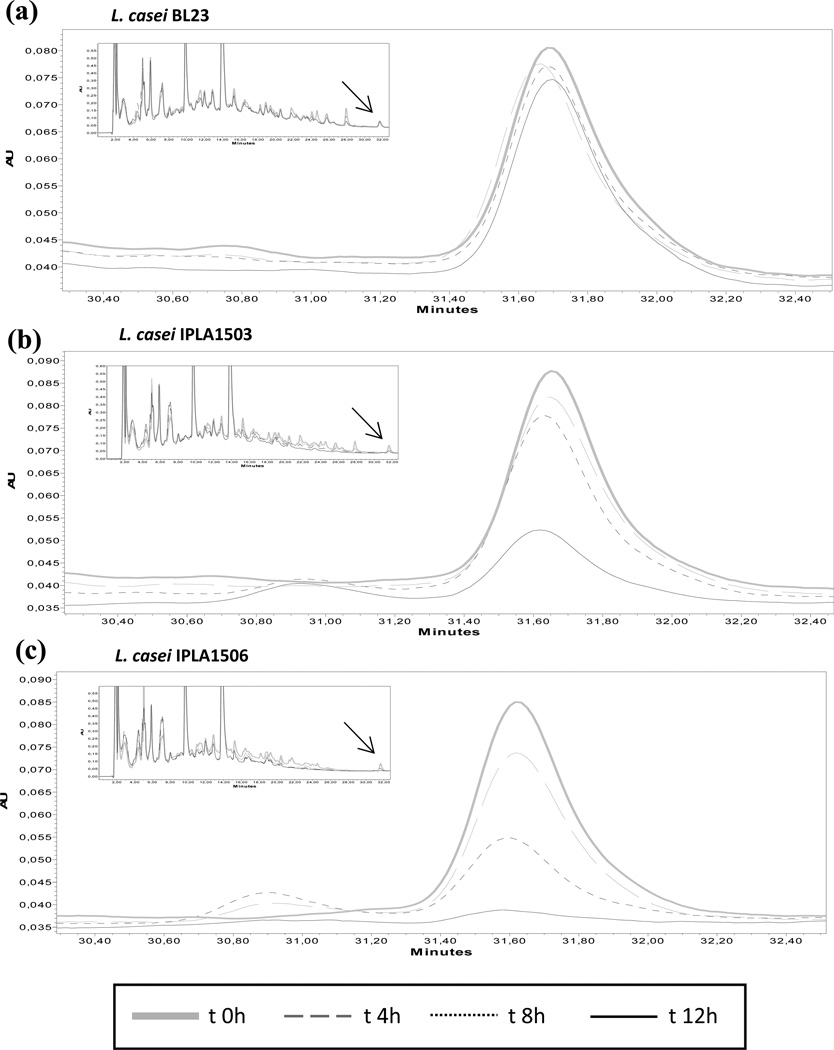
Fig. 5. Hydrolysis of the immunotoxic 33-mer peptide.
The strains L. casei BL23 (a), L. casei IPLA1503 (b) and L. casei IPLA1506 (c) were incubated with the 33-mer (25.5 µM) during 12 hours. Every four hours aliquots were removed and the substrate concentration was monitored by RP-HPLC analysis. Chromatograms show the hydrolysis time course of 33-mer peptide by the strains. The complete chromatogram appears in a box with the substrate arrowed.

