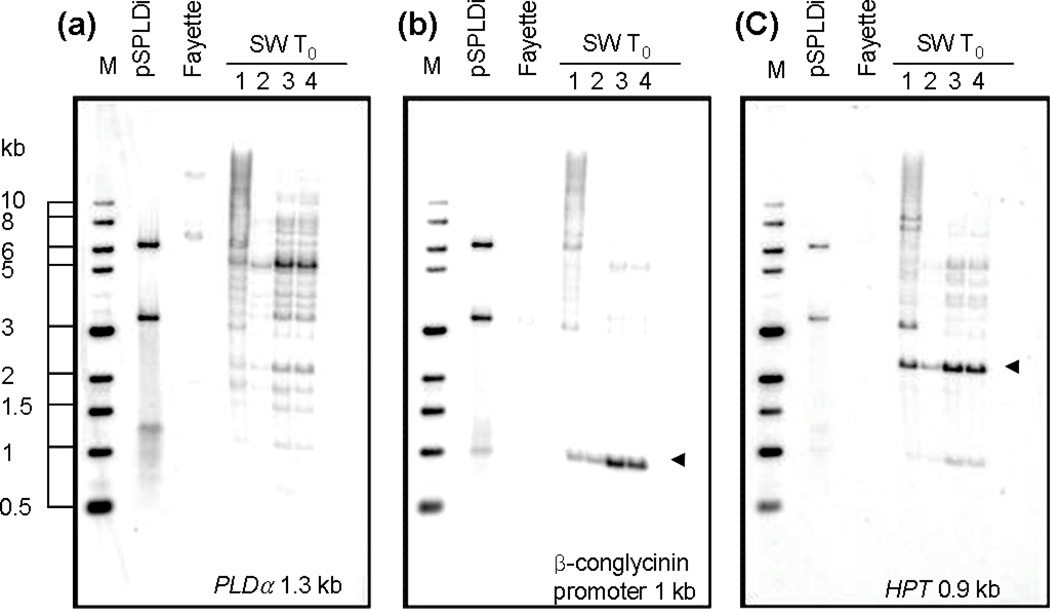
Figure 2.
Southern blot analysis of PLDα-suppressed T0 soybean lines with negative controls. Fifteen µg of genomic DNA were digested with HindIII. Digested genomic DNA from SW T0 young leaves was hybridized with a PLDα probe (a), beta-conglycinin promoter probe (b), and HPT probe (c). The probe used for DNA-DNA hybridization was indicated on the bottom of each gel image. pSPLDi transgene was linearized by HindIII digestion (a), whereas the β-conglycinin promoter (b) and HPT gene (c) were digested by HindIII. Triangles indicate the bands corresponding to the β-conglycinin promoter (b) and HPT gene (c) in the transgene in individual PLDα-suppressed T0 soybean, SW1 to 4.

