
Fig. 4.
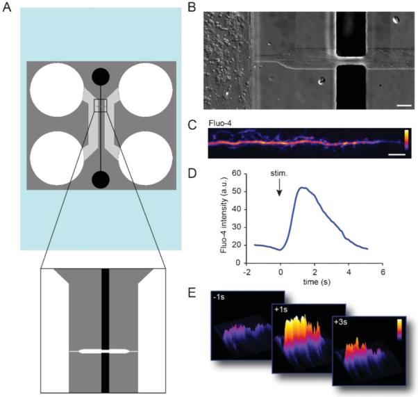
Liquid metal electrodes align to an axonal channel to depolarize axons. (A) Schematic of the axon-stim chamber. (B) Neuronal growth within the device. Axons are visible extending into the axonal channel in fluidic contact with the liquid metal electrodes. Scale, 50 μm. (C) An example of an axon labelled with Fluo-4 calcium indicator dye within the axon channel. Color lookup table, “Fire”. Scale bar, 20 μm. (D) Fluo-4 intensity within an axonal ROI over time. (E) Surface intensity maps at different time points for the axonal ROI.