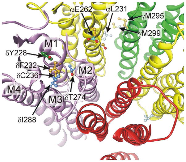
Figure 4. The δ-subunit four-helix bundle site.
The nAChR TMD structure is depicted as backbone ribbons, viewed from above. The δ subunit four-helix bundle is shown along with another perspective on the γ+/α-interfacial site. Anesthetic photolabeled residues are shown as ball-and-stick structures, colored according to which anesthetics modified them: halothane = green; azi-etomidate = gold; TID or aziPm = dark blue; azi-octanol = cyan.

