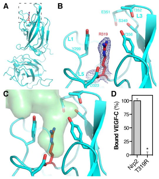
Figure 3. Crystal structure and VEGF-C binding properties of Nrp2-T319R.
(A) Structure of Nrp2-T319R with the stick representation for T319R shown in red. (B) Zoom of the Nrp2-T319R binding pocket. The blue mesh illustrates the 2Fo-Fc electron density map for R319 contoured at 1.0σ. (C) Superimposition of VEGF-C (green) onto the structure of Nrp2-T319R demonstrates that the binding pocket normally occupied by VEGF-C is blocked in the mutant. (D) VEGF-C binding was compared between Nrp2-b1b2 and Nrp2-T319R. Binding was measured in triplicate and is reported as mean ± SD (*p<0.05).

