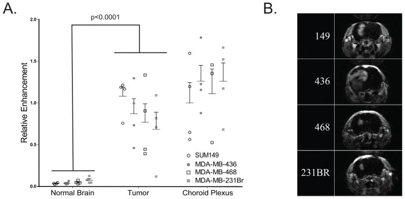
Figure 1. Blood brain barrier (BBB) permeability studies demonstrate compromised BBB in tumors as compared to normal brain and no significant differences in BBB permeability between models.
(A) Relative enhancement of four tumor cell lines with matched normal, tumor, and choroid plexus as determined from T1 DCE MR images, formed from digitally subtracting pre-contrast T1-DCE-weighted sequence from the identical sequence performed after gadolinium administration (20th sequence). Significant difference was observed between tumor and normal brain tissue (p<0.001), and no significant difference was seen between models within each tissue type (p>.14 within each tissue). (B) Brain MR imaging of a preresentative intracranial tumor from each of the four intracranial models.

