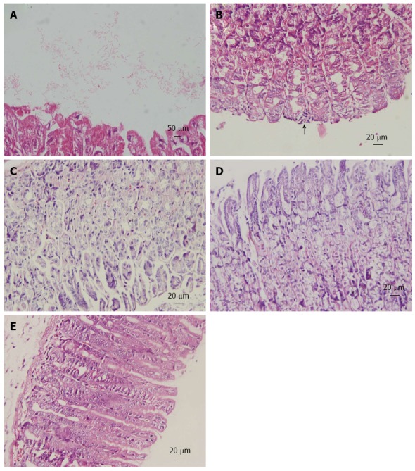
Figure 2.

Histopathology of gastric mucosa. Hematoxylin and eosin staining of gastric mucosa from mice inoculated with Helicobacter pylori. A: Helicobacter pylori (H. pylori) colonization on the surface of gastric mucosa (magnification × 100); B: Mononuclear cell infiltrates (arrow; magnification × 40); C: Uninfected mice exhibit a normal gastric mucosa (magnification × 40); D: Treatment with Chenopodium ambrosioides L. for 4 wk revealed no obvious inflammation (magnification × 40); E: Treatment with lansoprazole, metronidazole and clarithromycin for 1 wk showed no pathologic changes (magnification × 40).
