
Figure 3.
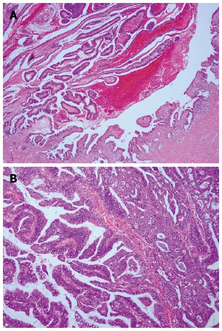
Histopathology of biliary tract intraductal papillary mucinous neoplasm. Hematoxylin and eosin staining of A: Common bile duct biliary tract intraductal papillary mucinous neoplasm, composed of papillary proliferation of atypical biliary epithelial cells (magnification × 40); and B: High-grade cytologic atypia and mucin in the numerous goblet cells (magnification × 100).