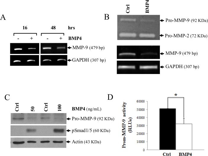
Figure 3. MMP-9 is decreased following BMP-4 stimulation in HT1080 cells.

(A) MMP-9 mRNA expression 24 and 48h following stimulation with recombinant BMP-4 (200 ng/ml). GAPDH was used as loading and specificity control. In (B), a zymogram (top gel) showing reduced MMP-9 secretion in supernatants of HT1080 cells treated for 16h with recombinant BMP-4 (200ng/ml). The lower panel shows the MMP-9 mRNA level of the treated cells. (C) Western blot analysis showing expression of MMP-9 and phosphorylation of Smad1/5 after treatment with human recombinant BMP-4. (D) Luciferase activity of in HT1080 cells transfected with a luciferase reporter vector containing the MMP-9-promoter following treatment with human recombinant BMP-4. Statistical analyses were carried out using Student's t test for unpaired samples. (* = p ≤ 0,05; ** = p ≤ 0,005).
