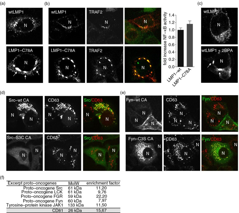
Fig. 3.
Palmitoylation controls subcellular trafficking of LMP1 but not TRAF2 association. (a) Immunofluorescent co-labelling of transfected wtLMP1 or LMP1-C78A (red) in HEK293 cells. N indicates nucleus. (b) Immunofluorescent labelling of endogenous TRAF2 (green) in HEK293 cells transfected with wtLMP1 or LMP1-C78A (red). To the right, reporter assay for LMP1-wt or LMP1-C78A NFκB activity. Cell lysates of HEK293 cells transfected for 24 hours with wtLMP1 or LMP1-C78A, together with an NFκB–reporter construct. Error bars represent s.e.m.; n>3. N indicates nucleus. (c) Immunofluorescent labelling of transfected wtLMP1 in 2BPA and control treated HEK293 cells. N indicates nucleus. (d, e) Immunofluorescent labelling of constitutive active Src-wt CA, Src-S3C CA, Fyn-wt CA, or Fyn-C3S CA (all in green) transfected HeLa-CIITA cells, co-labelled for CD63 (red). N indicates nucleus. (f) Table showing the enrichment of proto-oncogenes in exosomes versus cell lysates from 6 different B-cell lines referenced to the total number of peptides identified in each analysis.

