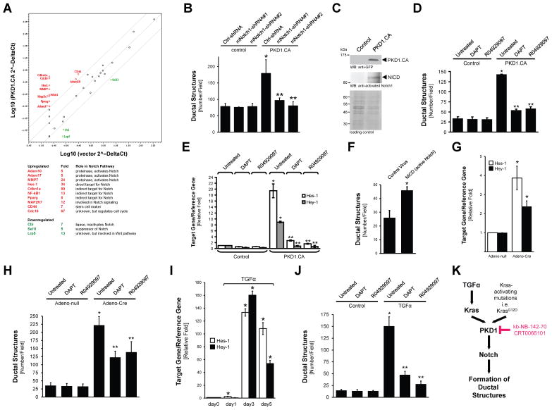
Figure 5. PKD1 mediates formation of duct-like structures through Notch.
(A) Scatter plot showing fold changes in expression of genes in control primary acinar cells or cells expressing constitutively-active PKD1 (PKD1.CA). The center line indicates no difference between the expression of genes under both conditions. The side lines indicate a 4-fold difference in either direction. The genes expressed more than 4-fold in the PKD1.CA group as compared to the control group are labeled in red. The genes expressed lower than 4-fold in the PKD1.CA group are labeled in green. Their role in the Notch pathway as well as fold change is provided in the table. (B) Primary acinar cells were isolated from mouse pancreas and infected with lentivirus harboring (scrambled) control shRNA (Ctrl-shRNA) or 2 different sequences of Notch1-shRNA (#1, #2) specifically targeting mouse Notch1. In addition cells were lentivirally-infected with active PKD1 (PKD1.CA) or control virus as indicated. Cells were then seeded in 3D culture in collagen and formation of ductal structures was quantified. (C) Primary acinar cells were isolated from mouse pancreas and lentivirally-infected with control or GFP-tagged active PKD1 (PKD1.CA; PKD1.S738E/S742E mutant). Cells were then seeded in 3D culture in collagen. At day 6, cells were re-isolated and analyzed by Western bot for activated Notch 1 fragment (NICD). Expression of PKD1.CA was controlled by Western blot against GFP. Silver staining served as a loading control. (D, E) After isolation and infection with control or PKD1.CA harboring lentivirus, primary acinar cells were seeded in collagen 3D culture in absence or presence of the γ-secretase inhibitors DAPT (5 μM) or R04929097 (10 nM) as indicated. At day 6, formation of ductal-structures was determined (D), or cells were isolated from the collagen and analyzed by quantitative PCR for expression of the Notch target genes Hes-1 and Hey-1 (E). (F) Primary acinar cells were isolated from mouse pancreas and infected with lentivirus harboring an active Notch fragment (NICD) or control virus. Cells were then seeded in 3D culture in collagen and formation of ductal structures was quantified. (G, H) Primary acinar cells from LSL-KrasG12D mice were isolated from mouse pancreas and expression of mutant Kras was induced by adenoviral infection of cre-recombinase as indicated (Adeno-Cre; or Adeno-null as control). Cells then were seeded in collagen 3D culture in absence or presence of the γ-secretase inhibitors DAPT (5 μM) or R04929097 (10 nM) as indicated. At day 6, cells were isolated from the collagen and analyzed by quantitative PCR for expression of the Notch target genes Hes-1 and Hey-1 (G), and formation of ductal-structures was determined (H). (I, J) Primary acinar cells were seeded in collagen 3D culture in absence or presence of TGFα (50 ng ml−1) and the γ-secretase inhibitors DAPT (5 μM) or R04929097 (10 nM) as indicated. At indicated days, cells were isolated from the collagen and analyzed by quantitative PCR for expression of the Notch target genes Hes-1 and Hey-1 (I), and formation of ductal-structures was determined (J). In all figures shown in B, D–J, * indicates statistical significance (p < 0.05; student’s t-test) as compared to control, ** indicates statistical significance (p < 0.05; student’s t-test) as compared to stimulus. Error bars (s. d.) were obtained from three experimental replicates. (K) Schematic of a TGFα- and Kras-induced PKD1-Notch signaling pathway that drives formation of pancreatic duct-like structures and PanIN formation. All experiments shown were performed at least 3 times with similar results.

