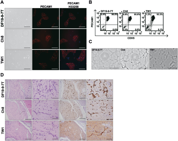
Figure 4. The validation of endothelial identity across multiple iPS/ES cell lines.
(A) Phase-contrast images (left panels) and immunofluorescence for PECAM1+ cells (middle and right panels, red) on day 5. Per well of a 12-well plate, iPS (DF19-9-7T) and ES (Ch8, and TW1) cells (20,000) were induced with MI for 48 hours and replated (30,000) in VM for additional 72 hours before staining for PECAM1 immunopositivity. H33258 served as the nuclear stain. Scale bars = 200 µm. (B) Representative flow cytometry for PECAM1+ and CDH5+ cells on day 5. iPS (DF19-9-7T) and ES (Ch8, and TW1) cells were induced as in A and harvested for staining with PE-conjugated anti-PECAM1 and FITC-conjugated anti-CDH5 on day 5. (C) Matrigel capillary assays for cell lines DF19-9-7T (left), Ch8 (middle), and TW1 (right). Per well of a 6-well plate, iPS/ES cells (40,000) were induced with MI for 48 hours and replated (60,000) in VM for additional 72 hours. On day 5, the cells were dissociated and seeded on Matrigel matrix (45,000/well in a 96-well plate) for the formation of capillary like-structures. Scale bars = 1 mm. (D) H&E (left panels) and anti-hPECAM1 immunohistochemical (right panels) staining of the injected Matrigel plugs. Per 10 cm dish, iPS/ES cells (300,000) were induced with MI for 48 hours and replated (900,000) in VM for additional 72 hours. The cells were dissociated and mixed with Matrigel matrix for subcutaneous injection in immunodeficient mice on day 5. The plugs were harvested 7 days after injection. Scale bars (low power) = 1 mm; scale bars (high power) = 200 µm.

