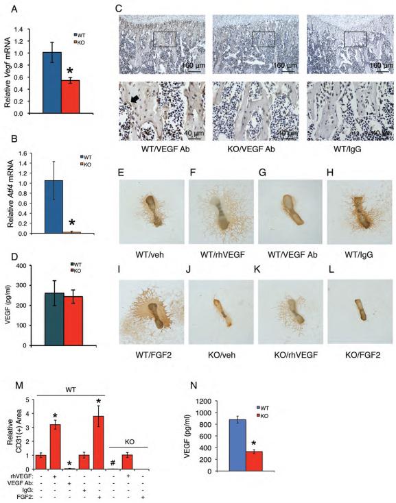
Figure 2. Loss of ATF4 reduces VEGF expression in osteoblasts located on trabecular bone surfaces and abolishes endothelial sprouting from Atf4−/− metatarsals, which is restored by exogenously supplied recombinant human VEGF.
(A and B) qPCR. Total RNAs were isolated from WT and Atf4−/− tibiae (n = 5-6) and analyzed by qPCR using specific primers for Vegf and Atf4 mRNAs, which were normalized to GapdhmRNA. *P < 0.01, versus WT. (C) IHC. Sections of WT and Atf4−/− tibiae (n = 5-6) were stained using specific antibodies against VEGF or normal IgG. VEGF signal was stained brown. Original magnification, x100 (top), x400 (bottom). (D) ELISA. Level of VEGF protein from WT and Atf4−/− serum was measured using an ELISA kit according to the manufacturer's instructions. n = 5-6. (E-M) Metatarsal angiogenesis assays. Metatarsals were dissected from WT and Atf4−/− E17.5 fetuses and cultured in α-MEM for 12 days, followed by IHC staining using anti-CD31 antibody as described in Materials and Methods. Representative images are shown. Microscope: Olympus SZ61. Original magnification, ×20. Endothelial sprouting from WT metatarsals (E) was significantly increased by the treatment of recombinant VEGF (10 ng/ml) (F) and blocked by an anti-VEGF neutralizing antibody (1 μg/ml) (G), but not by control IgG (1 μg/ml) (H). Endothelial sprouting from WT metatarsals was increased by FGF2 (100 ng/ml) (I). No detectable endothelial sprouting from Atf4−/− metatarsals (J). Significant endothelial sprouting from Atf4−/− metatarsals treated with VEGF (K), but not FGF2 (L). Quantitative data of each group (M). n = 6-8, *P < 0.01, versus veh, #P < 0.01, versus WT. (N)ELISA. Level of VEGF protein from WT and Atf4−/− metatarsal cultures was measured using an ELISA kit according to the manufacturer's instructions. n = 6-8, *P < 0.01, versus WT.

