Fig. 7.
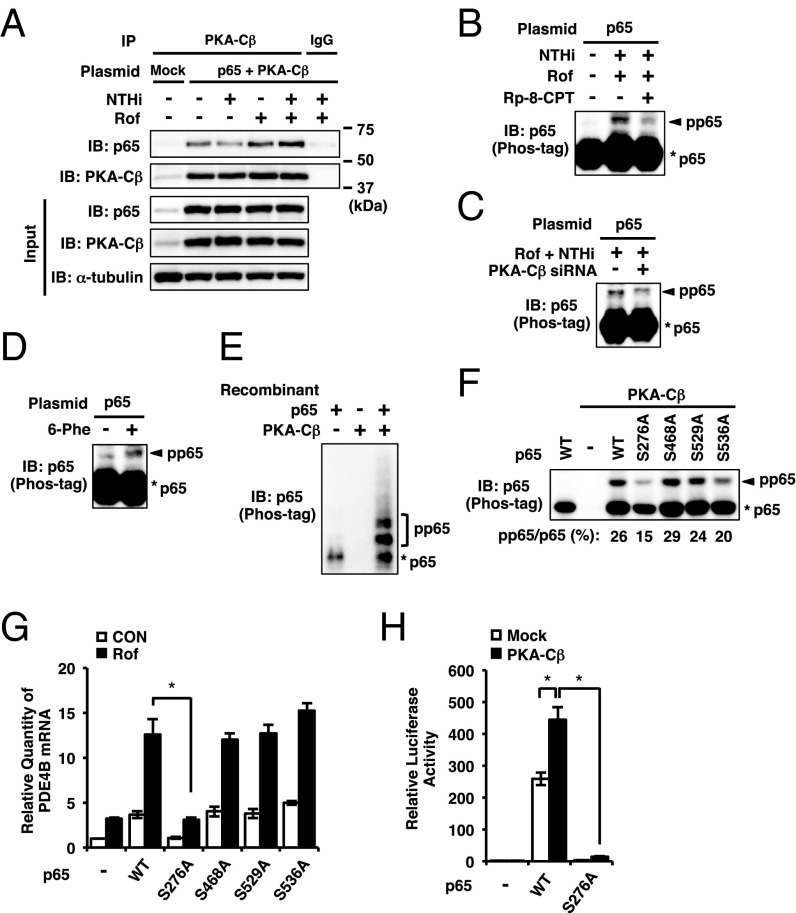
PKA-Cβ phosphorylates p65. (A) BEAS-2B cells were transfected with mock or p65 together with PKA-Cβ. After 48 h, cells were pretreated with Rof (0.1 µM) for 1 h followed by 1-h stimulation with NTHi. PKA-Cβ in whole-cell extracts was pulled down with anti-PKA-Cβ antibody and immunoblotted against p65 and PKA-Cβ. (B–D and F) BEAS-2B cells were transfected with plasmid or siRNA for 48 h, and p65 phosphorylation in nuclear extracts was analyzed by Phos-tag SDS-PAGE. Rp-8-CPT-cAMPS (Rp-8-CPT, 0.1 mM) was pretreated for 0.5 h before the Rof treatment (B). Rof (0.1 µM) was pretreated for 1 h followed by 1-h stimulation with NTHi (B and C). Cells were treated with 6-Phe-cAMP (6-Phe, 0.5 mM) for 2 h (D). (E) Recombinant p65 and PKA-Cβ proteins were mixed and incubated at 37 °C for 0.5 h and analyzed by Phos-tag SDS-PAGE. (G) After 48-h transfection of plasmid, BEAS-2B cells were treated with Rof (0.1 µM) for 2.5 h, and PDE4B mRNA expression was analyzed. (H) BEAS-2B cells were transfected with NF-κB luciferase plasmid together with mock, p65, S276A p65, and PKA-Cβ. After 24 h, NF-κB activity was measured. Data in G and H are mean ± SD (n = 3); *P < 0.05. Data are representative of three or more independent experiments. CON, control; pp65, phosphorylated p65.

