Fig. 1.
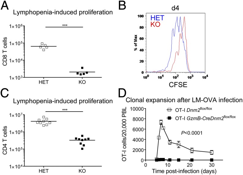
Dynamin 2 promotes T-cell proliferation in vivo. (A) Naive CD8 T cells from Dnm2 HET (CD45.1.2+) and KO (CD45.2+) mice were mixed 1:1 and injected into Rag1 KO recipients (CD45.1+). Number of CD8 T cells in spleen/lymph nodes recovered from Rag1 KO recipients 8 d after cotransfer is shown (n = 5). (B) Naive CD8 T cells from Dnm2 HET and KO mice were labeled with CSFE, mixed 1:1, and cotranferred into Rag1 KO mice. CFSE dilution was measured by flow cytometry 4 d after transfer. (C) Number of Dnm2 HET and KO CD4 T cells in spleen and lymph nodes recovered from Rag1 KO recipients 14 d after cotransfer (n = 9). A 1:1 mixture of naive Dnm2 HET and KO CD4 T cells was cotransferred into Rag1 KO recipients. (D) Number of Dnm2 WT and KO OT-I T cells in the blood of B6 recipient mice after infection with LM-OVA (n = 6–8). Naive CD8 T cells from OT-I Dnm2flox/flox (CD45.1.2+) and OT-I GzmB-creDnm2flox/flox (CD45.2+) mice were mixed 1:1 and injected into B6 recipients (CD45.1+). One day after transfer, recipient mice were infected with Listeria monocytogenes expressing OVA peptide (LM-OVA). All error bars represent SEM. ***P < 0.001 by unpaired Student's t test (A and C) or one-way ANOVA (D). Results are representative of or combined from two (A, B, and D), or three (C) experiments.

