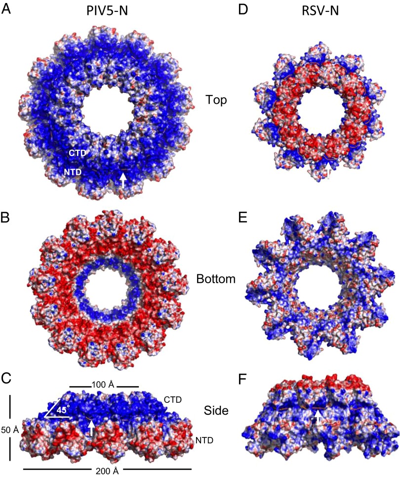
Fig. 3.
Molecular surface representation of PIV5-N (A–C) and RSV-N (D–F) nucleocapsid rings colored by the electrostatic potential, ranging from −10 kT/e (red) to +10 kT/e (blue). RNA strand (salmon) is indicated by white arrow for clarity. PIV5-N charge distribution is more polarized than RSV-N with positive charges on the outside-top of the ring and negative charges on the inside-bottom of the ring.

