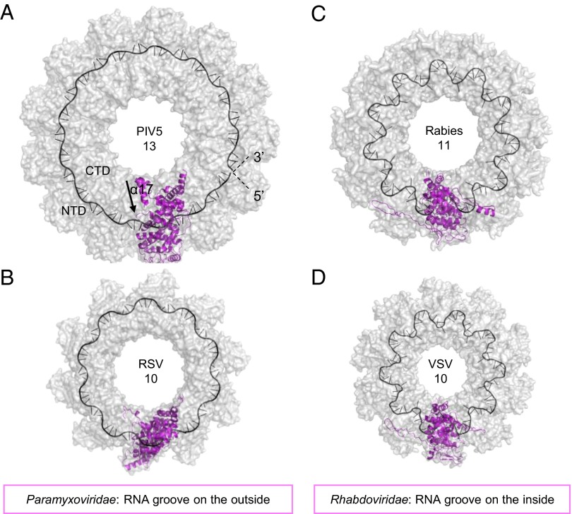
Fig. 4.
Surface representation (gray) of the nucleocapsid ring structures from different negative-strand RNA viruses. Paramyxoviridae virus nucleocapsid like PIV5-N 13-mer (A) and RSV 10-mer (B) with RNA (black cartoon) bound from the outside of the ring. Rhabdoviridae viruses like rabies 11-mer (C) and VSV 10-mer (D) with the RNA bound on the inside of the ring. One N-protomer in each ring is displayed as cartoon (magneta). Arrow in A indicates direction of helix α17.

