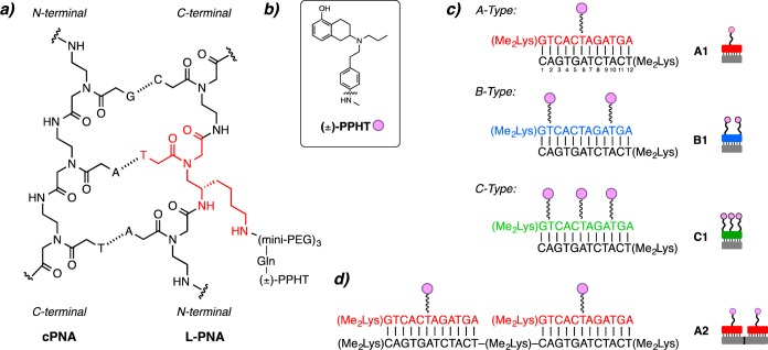
Figure 1.
Ligand modified PNAs. (a) Chemical structure of L-PNA:PNA duplex containing the lKγ-PNA side chain highlighted in red. (b) Chemical structure of D2R agonist (±)-PPHT (represented as pink circles). (c) L-PNA oligomer bound to complementary PNA with one (±)-PPHT ligand (A-type, red), two (±)-PPHT ligands (B-type, blue), and three (±)-PPHT ligands (C-type, green) per PNA. (d) Each L-PNA sequence is identified by its constituent parts; for example, an A2 complex contains 2 A-type L-PNA units annealed along a 24-residue cPNA.

